We reach more than 65,000 registered users in Dec!! Register Now
New fluorescent dye can light up the brain
- May 19, 2023
- 11 Views
- 0 Likes
- 0 Comment
Talk about a bright idea: Thanks to chemists at Rice University and Stanford University, lighting up the brain is no longer just a figure of speech.

“This could be very useful for imaging-guided surgery, for example,” Xiao said. “Using this dye, a doctor could determine where the boundary is between normal brain tissue versus tumor tissue.”
The study is featured on the cover of the Dec. 28 issue of the Journal of the American Chemical Society.
If you’ve been to an aquarium or a nightclub, you’ve probably noticed the colorful glow that some objects or surfaces emit under a black light. Known as fluorescence, this glowing effect can be useful for rendering visible things that otherwise go unnoticed.
“Fluorescence imaging has been applied for imaging cancer in different parts of our body,” Xiao said. “The advantages of a fluorescence probe include high resolution and the ability to adapt the probe to read for different substances or activities.”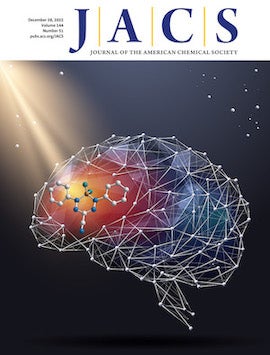
“Our tool is really valuable for deep imaging because it functions in the NIR-II region,” Xiao said. “In contrast to NIR-II wavelengths, fluorescent effects within the visible spectrum or with near-infrared wavelengths between 600 and 900 nanometers (NIR-I) will only get you skin-deep.”
Brain imaging poses a particular challenge not only because of tissue depth and accessibility, but also because of the blood-brain barrier, a layer of cells that acts as a very selective filter to restrict the passage of substances from the circulatory system to the central nervous system.
“People always want to know what exactly is happening in the brain, but it’s very hard to design a molecule that can penetrate the blood-brain barrier. Up to 98% of small-molecule drugs approved by the Food and Drug Administration (FDA) cannot,” Xiao said.
“Generally speaking, the reason a NIR-II dye molecule tends to be big is because it is a conjugated structure with many double bonds,” he continued. “This is a true problem and the reason why we have been unable to use fluorescence in brain imaging until now. We tried to address this issue by developing this new dye scaffold that is very small but has a long emission wavelength.”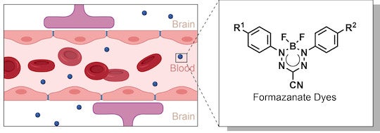
Beyond the brain, the dye developed by Xiao has much greater lasting power than indocyanine green, the only NIR small-molecule dye approved by the FDA for use as a contrast agent. A longer lifespan means researchers have more time to record the fluorescent trace before it disappears.
“When exposed to light, the indocyanine green dye trace deteriorates in seconds, whereas our dye leaves a stable trace for more than 10 minutes,” Xiao said.
List of Referenes
- Shichao Wang, Hui Shi, Lushun Wang, Axel Loredo, Sergei M. Bachilo, William Wu, Zeru Tian, Yuda Chen, R. Bruce Weisman, Xuanjun Zhang, Zhen Cheng, Han Xiao. Photostable Small-Molecule NIR-II Fluorescent Scaffolds that Cross the Blood–Brain Barrier for Noninvasive Brain Imaging. Journal of the American Chemical Society, 2022; 144 (51): 23668 DOI: 10.1021/jacs.2c11223
Cite This Article as
No tags found for this post