We reach more than 65,000 registered users in Dec!! Register Now

Cellular waste removal differs according to cell type
- March 17, 2023
- 30 Views
- 0 Likes
- 0 Comment
Study by the University of Bonn identifies different types of so-called lysosomes
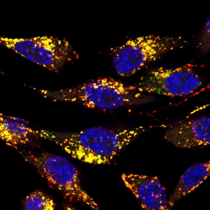
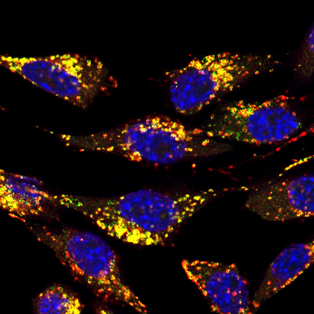
"Miniature shredders" are at work in each cell, disassembling and recycling cell components that are defective or no longer required. The exact structure of these shredders differs from cell type to cell type, a study by the University of Bonn now shows. For example, cancer cells have a special variant that can supply them particularly effectively with building blocks for their energy metabolism. The results were published online in advance. The final version has now been published in the journal "Molecular & Cellular Proteomics." Mouse skin cells - with nucleus stained blue. Lysosomes were stained red, and a newly discovered lysosomal protein was stained green. Where both occur together, this results in a yellow coloration.© Image: Florian Bleibaum/University of KielDownload all images in original sizeThe impression in connection with the service is free, while the image specified author is mentioned.Lysosomes are a central part of the cell's waste disposal system. The tiny bubbles, surrounded by a fat-like membrane, function like a miniature recycling factory: They break down defective cell components, harmful molecules, or proteins that are no longer needed into their individual parts. They then make these available to the cell again. "The process is extremely important," stresses Dr. Dominic Winter of the Institute of Biochemistry and Molecular Biology at the University Hospital Bonn. "If it doesn't work properly, diseases like Alzheimer's or Parkinson's can result."
Lysosomes have a very complex structure. Several hundred proteins are now known to play a role in their function. There could even be significantly more: When lysosomes are isolated from cells and their composition is analyzed with special equipment, researchers often find more than 5,000 different cellular proteins. "However, it's impossible to say how many of them are actually important for the lysosomes’ work," Winter says. "They can also be molecules that are in the process of being broken down in them. Others may be attached to their membrane from the outside, without performing any task. And there's usually a lot of unwanted bycatch when isolating the lysosomes, too."
100 new potential lysosomal proteins discovered
The researchers have developed a method that allows them to identify a large proportion of these uninvolved molecules. Of the 5,000 proteins typically found using conventional methods, this left around 1,000. "We performed this step for six very different cell types, including, for instance, liver cells and cancer cells," the researcher explains. "Several hundred of these 1,000 proteins were present in almost all lysosomes - no matter which tissue they originated from. These included about 100 new lysosomal proteins in addition to those already known. We believe it is likely that these also play an important role in the function of the nano-shredders."
What differed from cell type to cell type was the quantity in which each of these proteins was present. "The lysosomes of liver cells, for example, are packed to the brim with degradation enzymes," says Winter, who is also a member of the Transdisciplinary Research Area "Life & Health" at the University of Bonn. "And that makes sense - an important function of the liver is to break down different molecules. In contrast, in the cancer cells we studied, the lysosomes contained a lot of transporter proteins."
Tumors require a lot of energy for their growth; at the same time, they are often poorly supplied with blood. They therefore "digest" the surrounding tissue and use the breakdown products to obtain energy. Digestion takes place in the lysosomes, which then have to transport the broken-down molecules back into the cell - hence the many transporters. This means that these nano-shredders differ according to the requirements of the tissue in which they are found. "In each of the six cell types we studied, the lysosomes have a very specific protein makeup," Winter points out. "To my knowledge, we are the first research group that has been able to show that."
Protein fingerprint provides clues to disease mechanisms
On the one hand, the results are particularly interesting for basic research. On the other hand, they also shed new light on the role of lysosomes in certain diseases. There are more than 1,600 studies worldwide on a wide variety of disorders suggesting the involvement of lysosomal proteins. For example, it has been known for some time that the lysosomes of very specific nerve cells are altered in Parkinson's disease. "We can now take a kind of protein fingerprint of these lysosomes and compare it to that of healthy individuals," Winter explains. "This could provide clues to how the cellular shredder function is altered in affected individuals and why that causes neurological problems." In the long term, this could also help find new approaches for drugs.
Although the study was conducted at the University Hospital Bonn, it is ultimately also a multinational project: The three lead authors are from three different countries - DAAD scholarship holder Dr. Fatema Akter from Bangladesh, her colleague Srigayatri Ponnaiyan from India, and doctoral student Sara Bonini from Italy.
List of Referenes
- Fatema Akter, Sara Bonini, Srigayatri Ponnaiyan, Bianca Kögler-Mohrbacher, Florian Bleibaum, Markus Damme, Bernhard Y. Renard, Dominic Winter. Multi–Cell Line Analysis of Lysosomal Proteomes Reveals Unique Features and Novel Lysosomal Proteins. Molecular & Cellular Proteomics, 2023; 22 (3): 100509 DOI: 10.1016/j.mcpro.2023.100509
Cite This Article as
No tags found for this post


