


We reach more than 65,000 registered users in Dec!! Register Now
WEARABLE MICROSCOPES ADVANCE SPINAL CORD IMAGING IN MICE
- March 30, 2023
- 51 Views
- 0 Likes
- 0 Comment
Salk scientists invent wearable microscopes to produce high-definition, real-time images of mouse spinal cord activity across previously inaccessible regions
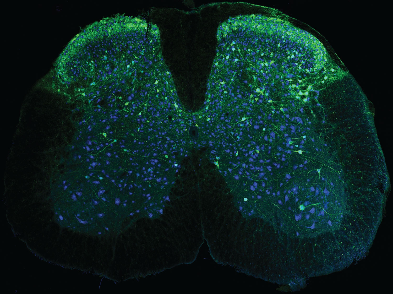
The spinal cord acts as a messenger, carrying signals between the brain and body to regulate everything from breathing to movement. While the spinal cord is known to play an essential role in relaying pain signals, technology has limited scientists’ understanding of how this process occurs on a cellular level. Now, Salk scientists have created wearable microscopes to enable unprecedented insight into the signaling patterns that occur within the spinal cords of mice.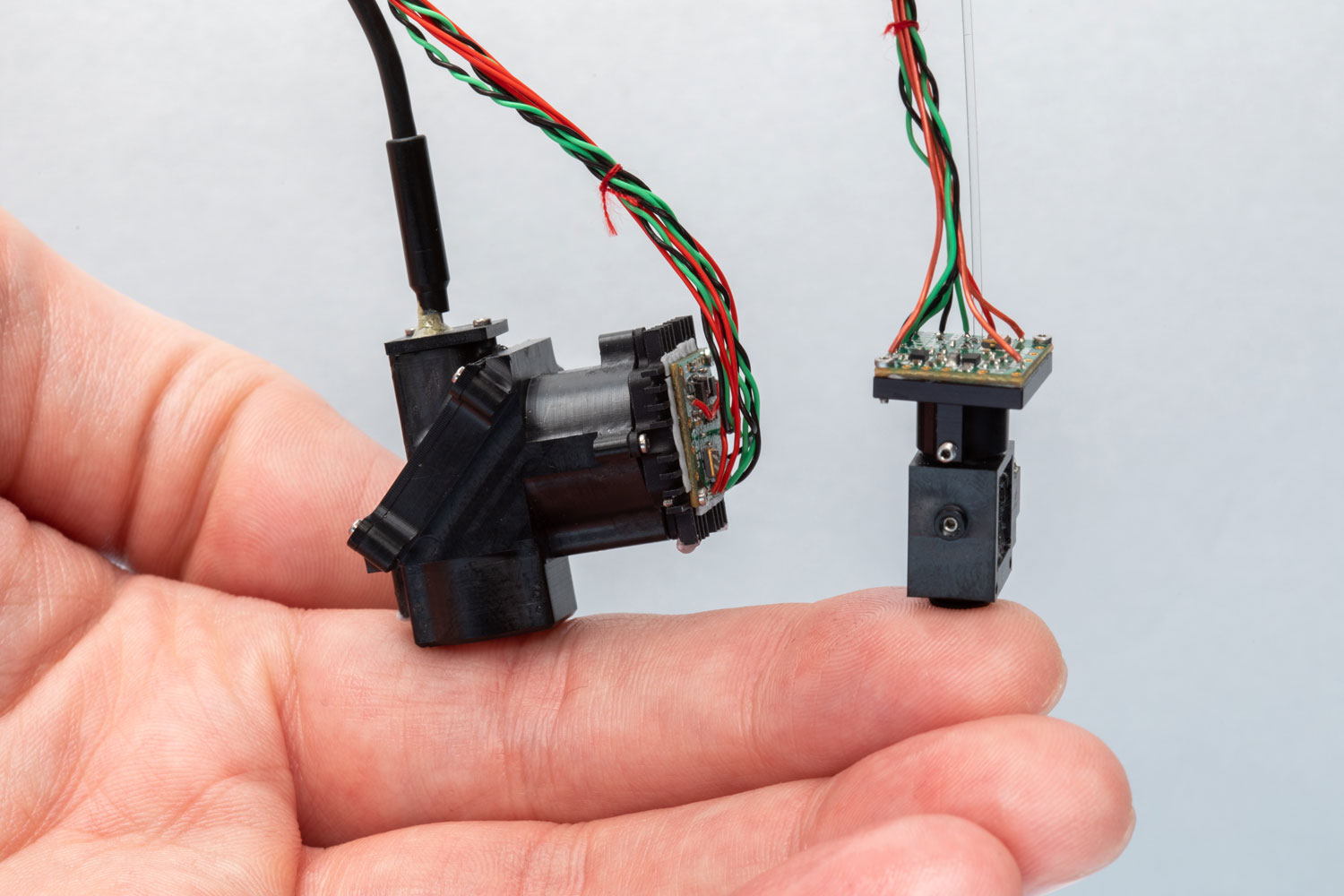
“These new wearable microscopes allow us to see nerve activity related to sensations and movement in regions and at speeds inaccessible by other high-resolution technology,” says senior author Axel Nimmerjahn, associate professor and director of the Waitt Advanced Biophotonics Center. “Our wearable microscopes fundamentally change what is possible when studying the central nervous system.”
The wearable microscopes are approximately seven- and fourteen- millimeters wide (about the width of a little finger or the human spinal cord) and offer high-resolution, high-contrast, and multicolor imaging in real-time across previously inaccessible regions of the spinal cord. The new technology can be combined with a microprism implant, which is a small reflective glass element placed near the tissue regions of interest.
Pavel Shekhtmeyster, a former postdoctoral fellow in Nimmerjahn’s lab and co-first author on both studies, agrees, “We’ve overcome field-of-view and depth barriers in the context of spinal cord research. Our wearable microscopes are light enough to be carried by mice and allow measurements previously thought impossible.”
With the novel microscopes, Nimmerjahn’s team began applying the technology to gather new information about the central nervous system. In particular, they wanted to image astrocytes, star-shaped non-neuronal glial cells, in the spinal cord because the team’s earlier work suggested the cells’ unexpected involvement in pain processing.
The team found that squeezing the tails of mice activated the astrocytes, sending coordinated signals across spinal cord segments. Prior to the invention of the new microscopes, it was impossible to know what astrocyte activity looked like—or what any cellular activity looked like across those spinal cord regions of moving animals.
Nimmerjahn’s team has already begun investigating how neuronal and non-neuronal activity in the spinal cord is altered in different pain conditions and how various treatments control abnormal cell activity.
Other authors include Alexander Ngo, Grace Gao, Nicholas A. Nelson, Jack A. Olmstead, and Charles L. Clark of Salk.
The work was supported by the National Institutes of Health (R01NS108034, U19NS112959, U19NS123719, U01NS103522, and F31NS120619), a National Institutes of Health Training Grant (T32/CMG), the Sol Goldman Charitable Trust, C. and L. Greenfield, a Rose Hills Foundation Graduate Fellowship, a Burt and Ethel Aginsky Research Scholar Award, a Kavli-Helinski Endowment Graduate Fellowship, and a Salk Innovation Grant.
List of Referenes
- Pavel Shekhtmeyster, Erin M. Carey, Daniela Duarte, Alexander Ngo, Grace Gao, Nicholas A. Nelson, Charles L. Clark, Axel Nimmerjahn. Multiplex translaminar imaging in the spinal cord of behaving mice. Nature Communications, 2023; 14 (1) DOI: 10.1038/s41467-023-36959-2
Cite This Article as
No tags found for this post

